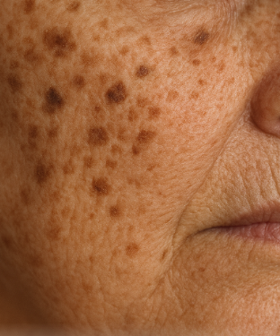
Liver spots, more properly called solar lentigines or age spots, are among the most visible signs that skin carries a history. They appear as flat, well-defined patches of pigment on areas habitually exposed to sunlight and are a visual ledger of cumulative ultraviolet exposure and biological aging. These marks are cosmetic rather than systemic in origin, but their presence speaks to layered causes—environmental, behavioral and biological—that intersect over decades to produce the familiar brown macules on the face, hands and décolletage. The list that follows parses the ten most common contributors to liver spots, explaining the mechanisms by which each factor influences pigmentation and offering journalistic clarity about prevalence and context while carefully avoiding medical advice.
1. Cumulative Ultraviolet Exposure
Ultraviolet (UV) radiation is the principal architect of liver spots. Repeated exposure to the sun’s UV rays stimulates melanocytes—the pigment-producing cells—to increase melanin production and, over time, to cluster in localized areas. The result is the discrete, darker patches clinicians and consumers call liver spots. These lesions are most common on the face, the backs of the hands, shoulders and forearms—sites of chronic sun exposure—and they typically become more pronounced with advancing age as cumulative damage accrues. Tanning beds, which emit concentrated UVA and UVB radiation, operate by the same biological logic and therefore pose comparable risk for accelerating the formation of solar lentigines.
2. Age-Related Changes in Skin Biology
Aging skin is less adept at maintaining uniform pigment distribution. Cell turnover slows, DNA repair capacity weakens and the epidermal-dermal interface undergoes structural changes that together make irregular pigment deposition more likely. The combination of diminished regenerative rhythm and accumulated photodamage increases the visibility and persistence of lentigines as people pass middle age and beyond. Epidemiological reporting consistently shows a higher prevalence of liver spots in older adults, reflecting both cumulative environmental exposure and intrinsic aging processes.
3. Genetic Predisposition
Not everyone with substantial sun exposure develops pronounced liver spots, and heredity helps explain this variability. Genetic factors influence baseline melanocyte reactivity, the density and distribution of pigment cells, and the skin’s structural resilience. Family histories of early-onset or particularly numerous age spots suggest inherited susceptibility—an amplification of risk rather than a deterministic path. Genetics operates as a background amplifier: given equal UV exposure, some individuals will exhibit more pronounced lentigines because their skin’s response to photic stimuli is innately different.
4. Fair Skin and Lower Baseline Melanin
Skin phototype matters. Individuals with lighter skin tones possess less baseline melanin, which provides less intrinsic UV shielding; consequently, their melanocytes are more likely to overcompensate with focal hyperpigmentation after repeated UV insult. Liver spots are therefore statistically more common and often more noticeable in fair-skinned populations. That said, they are not exclusive to one skin type; people with medium and darker complexions can and do develop lentigines, though the clinical appearance and contrast with surrounding skin will differ by pigmentary context.
5. History of Sunburns and Intermittent Intense Exposure
There is a qualitative difference between daily, moderate sun exposure and episodic, intense UV events such as sunburns. Acute, high-dose exposure causes cellular injury and inflammatory signaling that can precipitate irregular melanin deposition. A life history punctuated by sunburns—particularly in youth—creates a pattern of accumulated, episodic damage that often manifests as uneven pigmentation decades later. Epidemiological studies associate repeated sunburns with an increased likelihood of later-life photodamage, including lentigines and other manifestations of photoaging.
6. Hormonal Influences and Photosensitizing Agents
Hormones modulate pigmentation. While liver spots are primarily driven by UV and age, hormone-related pigmentation conditions—such as melasma—demonstrate the skin’s sensitivity to endocrine signals. Moreover, certain medications and topical agents increase photoreactivity; photosensitizing drugs or cosmetic actives can make skin more responsive to UV exposure, raising the odds that pigment will accumulate in discrete patches. The interaction is not straightforward: hormones and photosensitizers do not cause lentigines wholesale, but they can alter the skin’s reactivity to UV triggers, shifting risk in clinically meaningful ways.
7. Cumulative Photodamage From Environmental UV Sources
Beyond intentional sun exposure, everyday environmental UV—reflected light, incidental midday exposure and occupational outdoor time—contributes to cumulative burden. Urban living, with reflective glass, concrete and incidental midday exposure during commutes or lunchtime, can incrementally increase photodamage compared with a life lived predominantly indoors. The aggregate effect across years explains why liver spots often appear in midlife: they are the composite record of incidental and intentional UV interactions that slowly remodel pigment distribution.
8. Tanning Bed Use and Artificial UV Exposure
Tanning beds accelerate a process that outdoor sun does more slowly; they deliver concentrated UVA and UVB radiation that increases melanin production and damages cutaneous DNA and supporting structures. Use of artificial tanning devices has been linked to a range of photodamage outcomes, including earlier and more pronounced pigmentation changes, because the intensity and spectrum of radiation in tanning beds can be greater than many natural exposures. The mechanism—UV-induced melanocyte activation and damage-mediated dysregulation of pigment distribution—is the same, but the tempo of change is often faster with artificial sources.
9. Photoaging-Related Structural Changes in the Dermis
Photoaging describes the structural degradation of the dermis from chronic UV radiation: collagen fragmentation, elastin accumulation, and alterations in the extracellular matrix that together change the skin’s mechanical and optical properties. These dermal shifts can make epidermal pigment irregularities more apparent; the same amount of melanin appears different when the skin’s microarchitecture is altered. In effect, photoaging makes lentigines both more likely to develop and more visually prominent when they do, because changes in dermal scattering and epidermal thickness modulate how pigment is perceived at the surface.
10. Cumulative Lifestyle and Occupational Factors
Finally, lifestyle and occupation shape cumulative exposure. Outdoor labor, sports, long commutes, and leisure patterns that prioritize midday sun increase lifetimes of UV interaction. Clothing choices and inconsistent sun protection habits—intermittent use of protective clothing, hats or sunscreen—compound the risk. Occupational exposures that combine UV with mechanical friction or chemical sensitizers can create local environments more conducive to dysregulated pigmentation. The practical upshot: similar to the way personal style accumulates into a visible aesthetic, years of habitual exposure and protection choices leave a pigmentary signature on the skin.
Putting the Causes in Context
The causes listed above do not act in isolation. Instead, they form a lattice of risk where intrinsic factors—age, genetics, baseline phototype—set a predispositional tone and extrinsic factors—UV exposure, tanning devices, occupational and lifestyle variables—determine the degree and distribution of visible damage. Scientific summaries and clinical reviews underscore that solar lentigines are primarily a photodamage phenomenon: melanin’s unevenness is a response to cumulative UV-mediated injury in an aging epidermis and dermis. That causal architecture explains why the condition is common after middle age and why prevention strategies emphasized in public-health literature focus on limiting UV dose throughout life.
Cultural and Cosmetic Perspectives
In fashion and beauty spheres, liver spots occupy a fraught terrain between stigma and acceptance. Historically hidden or treated as imperfections to be erased, they increasingly appear in editorial spreads and campaigns that aim to present more varied skin narratives. Yet the cosmetic impulse to lighten or remove age spots—through topical agents, in-office procedures or camouflage—remains robust, fueled by aesthetic norms and by the commercial interests of skincare and dermatology sectors. The tension is not merely stylistic: it reflects broader questions about how culture values signs of age, how commercial markets respond, and how individuals navigate choices about appearance that are deeply personal and materially consequential.
Evidence, Limitations and the Need for Clinical Discrimination
The evidence base for what causes liver spots is robust on the UV and aging front, and large-scale epidemiology and clinical review consistently emphasize photodamage as the dominant driver. However, not every dark patch is a benign lentigo; irregular or changing pigmentation warrants clinical evaluation to exclude lentigo maligna, melanoma or other pigmented lesions. The journalistic role is to synthesize population-level knowledge. Clinical discrimination and histopathological assessment remain the province of qualified practitioners when a spot is atypical in color, border or evolution. Ensure you schedule a time to see
Conclusion
Liver spots are a visible archive of the skin’s encounter with time and light. Age, genetics and baseline phototype shape susceptibility, while cumulative UV exposure—both intentional and incidental—drives the pigmentary changes that clinicians label as solar lentigines. Hormonal factors, photosensitizing agents, tanning-bed use and the structural consequences of photoaging modulate how and when these spots appear, and lifestyle or occupational exposure patterns contribute to an individual’s pigmentary biography. Understanding these causes illuminates why liver spots are so common in midlife and older adults and why public-health messages that reduce lifetime UV burden are so central to changing the population-level trajectory of visible photodamage.
